This dataset is designed to have the most reliable ground truth myocardial contours from short-axis MRI. We have collected not too many cases (only 15) from different vendors (3 MR systems) with multiple pathologies (1 healthy and 3 cardiac disease). We then asked 7 top MR core-labs from the Society of Cardiovascular Magnetic Resonance to manually draw myocardial contours by their expert reader. No restriction on how to draw these contours. We simply asked them to use the standard clinical protocol for MRI analysis applied in their labs. We derived the consensus contours from their manual contours.
You can read more information about this project from [Suinesiaputra et al., JCMR 2015].
Performance report
When you submit your contours, we will process your contour files and provide you with the following reports:
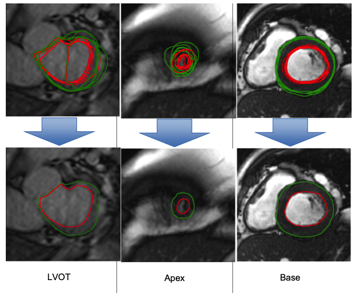
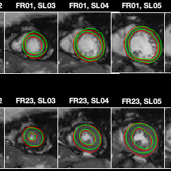
Contour Snapshots
Plots of your contours and the consensus contours for each image.
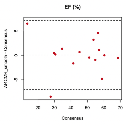
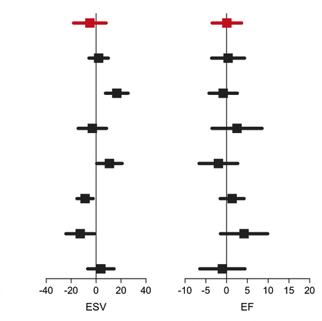
Bland-Altman Plots
Comparisons of EDV, ESV, LV mass and ejection fraction.
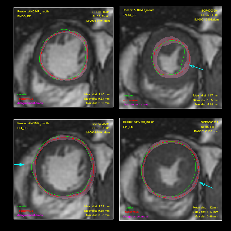
Disagreement Plots
Disagreement areas with the consensus contours for each contour.
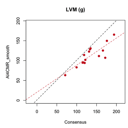
Scatter Plots
Scatter plots of EDV, ESV, LV mass and Ejection Fraction.
Reader Biases
Disagreement areas with the consensus contours for each contour.
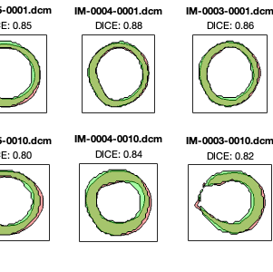
Intersection Plots
Intersection areas to visually show Dice coefficients.
Myocardial Area
Spreadsheet of endocardium and epicardium areas for each image.
Dice Coefficients
Spreadsheet of Dice coefficients for each image.
LV Functions
Spreadsheet of EDV, ESV, EF, LVM and epicardial volumes.
For submission, please contact us by email.
Contributors
- Prof. Dr Eike Nagel - University Hospital Frankfurt am Main, Germany
- Prof. Alistair Young - King's College London, United Kingdom
This consensus contour dataset project has been endorsed and supported by the Society of Cardiovascular Magnetic Resonance.